Our Technology
Background
Oscillution expertise and capabilities dates back to 2006, when Oscillutions founder pioneered the use of stem cell-derived brain cells with microelectrode arrays to profile the effects of compounds on neuronal network function in vitro.
From immaturity to functionality
Our technology is based on four key in-house expertise:
- differentiation of healthy and patient–derived human iPSC into 3D in vitro models of human brain cells (cortical neurons, astrocytes and oligodendroglial cells)
- combining pluripotent stem cell-derived brain cells with 96-well microelectrode-array plates.
- analyzing neuronal network (spiking and population bursting) and oscillatory activity (local field potentials 0.1-200Hz) measured from human brain cells.
- revealing compound effects on the full spectra of replicated human brain functionality.
Replicating human brain cellular composition
We differentiate human iPSC towards 3D brain aggregates of human astrocytes, oligodendroglia cells and neurons with electrophysiological functionality as recorded from brain tissue

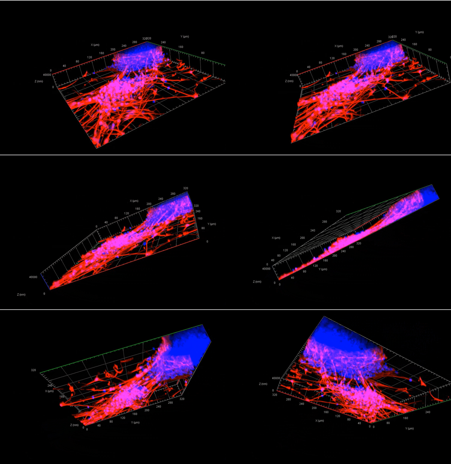
3D image reconstruction of adherent 3D brain aggregates shows human iPSC-derived neurons stained with beta-tubulin III (red) and DAPI-stained nuclei (blue).

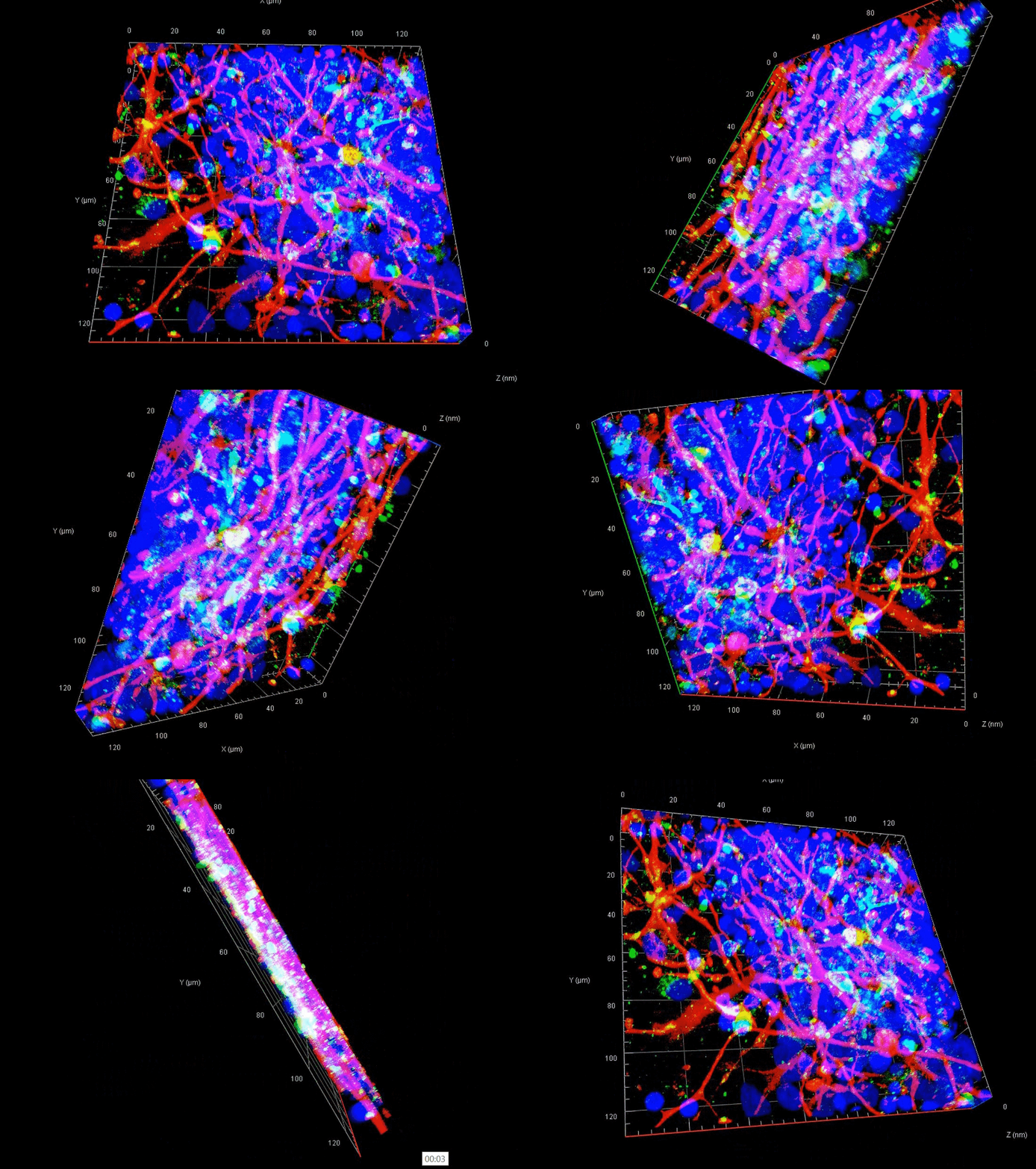
3D image reconstruction shows human iPSC-derived GFAP+ astrocytes (red), O4+ oligodendroglial cells (green), and DAPI-stained nuclei (blue) within adherent 3D brain aggregates.
Replicating human brain functionality at high capacity
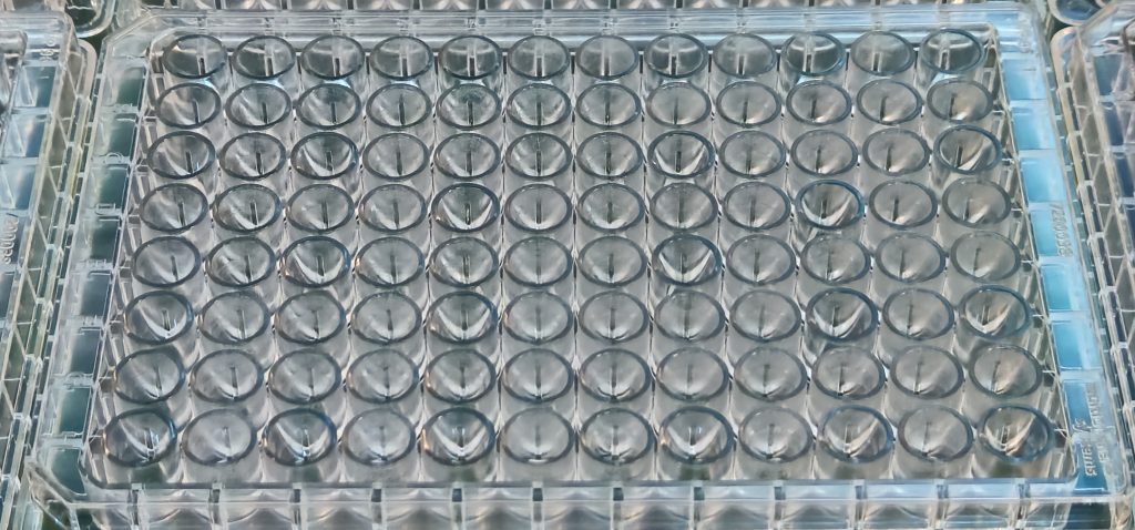
For analyzing human brain functionality in vitro at high-capacity, we utilize 96-well plates with microelectrode arrays within each well to record the full spectrum of electrical signals generated from human iPSC-brain cells we make.
Our proprietary software is continuously enhanced to accurately extract and analyze these signals, capturing spikes, bursts, network bursts and local field potentials across slow-oscillation, delta, theta, alpha, beta, gamma, and high-frequency domains.

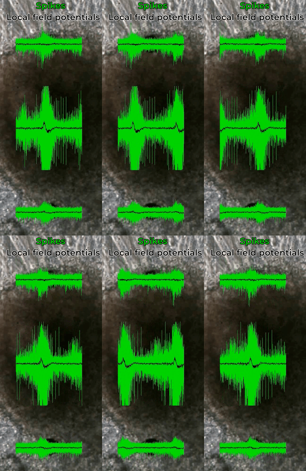
The video shows the spiking (green) and local field potential (black) activity recorded from 3D brain aggregates adherent to a microelectrode array.

